What is Digital Infrared Thermal Imaging?
Digital Infrared Thermal Imaging, or DITI, is a totally non-invasive, painless procedure with no radiation and no contact with the body.
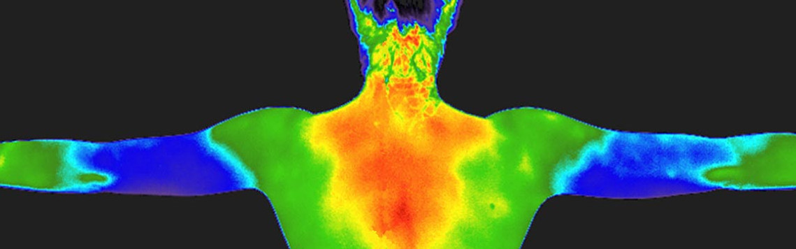
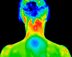
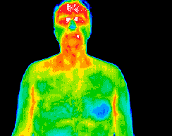
It uses an infrared camera with a highly accurate heat sensor to detect differences in temperature in the body. The body’s temperature is displayed graphically in different colours on a screen. Normally, the body’s temperature is symmetrical but abnormalities, injuries and pathologies change the temperature of the surrounding tissue and these differences are clearly visible on the scan.
Digital Infrared Thermal Imaging can help you with
- To help in determining the cause of pain.
- To aid in the early detection of disease and pathology.
- To evaluate sensory-nerve irritation or significant soft-tissue injury.
- To define a previously diagnosed injury or condition.
- To identify an abnormal area for further diagnostic testing.
- To follow progress of healing and rehabilitation.
How does Digital Infrared Thermal Imaging work?
Skin blood flow is under the control of the sympathetic nervous system. In normal people there is a symmetrical pattern across the skin which is consistent and reproducible for any individual. Our DITI scanner records this temperature pattern at a sensitivity of 0.01°C. The pattern is represented graphically by different colours on your thermograph or image.
Injuries, pathologies and tumours show up as ‘hot’ areas (or sometimes abnormally cold areas) in comparison to your normal temperature pattern. Vascular conditions such as Raynauds, Vasculitis, Limb Ischemia, Deep Vein Thrombosis also show up on DITI scans.
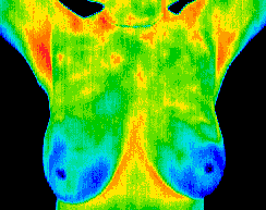
Normal breast image.
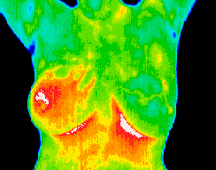
Inflammatory carcinoma.
Pregnancy.
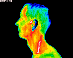
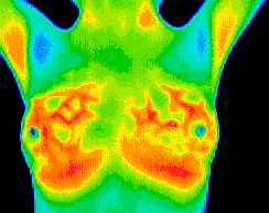
Carotid artery inflammation.
Underactive thymus gland = chronic fatigue syndrome.
Dental infection/cardiovascular dysfunction.
Digital Infrared Thermal Imaging can help with...
- Early detection of breast disease including cancer, fibrocystic disease, infection and avascular disease
- Diagnosis of neuromusculoskeletal injuries in sports people and their prognosis for return to participation and/or competition
- Diagnosis of dental disease, prior to feeling any pain
- Stroke screening
- Evaluating injuries and monitoring the healing process
- Detection of Deep Vein Thrombosis
Digital Infrared Thermal Imaging FAQs
What happens during a scan?
This quick and easy test starts with your medical history being taken before you partially disrobe for the scanning to be performed. This first session provides
the baseline of your “thermal signature”. A subsequent session assures that the patterns remain unchanged. All of your thermograms (images) are kept on record and
once your stable thermal pattern has been established any changes can be detected during your routine annual studies.
Digital Infrared Thermal Imaging research
There is a growing body of research supporting the use of thermographic imaging for early diagnosis of abnormalities and monitoring of healing. Below is just a small sample or Click Here to download this fuller selection of research.
Gautherie, M., et al. (1983). Thermobiological assessment of benign and malignant breast diseases. American Journal of Obstetrics & Gynecology, (8)147, pp.861-869.
“This study analysed the survival rates of 106 patients in whom the diagnosis of breast cancer was established as a result of the follow-up of thermographic
abnormalities found on the initial examination when the breasts were apparently healthy (negative physical and mammographic findings). A 61% increase in survival
was noted in the patients who were followed-up due to initial thermographic abnormalities. The authors summarised the study by stating that “the findings clearly
establish that the early identification of women at high risk of breast cancer based on the objective thermal assessment of breast health results in a dramatic
survival benefit.”
Gautherie, M. & Gros, C.M. (1980). Breast thermography and cancer risk prediction. Cancer, 45, pp. 51-56.
“Thermography is a useful predictor of risk factor for cancer and as an assessment tool for rapidly growing tumours.”
Wladisalw, V. E. et al. (1989). Screening thermography of chronic back pain patients with negative neuromusculoskeletal findings. Thermology, 3, pp. 125-126.
“This blind study indicated that thermograph imaging is effective at detecting neuromusculoskeletal abnormalities.”
Varju, G. et al. (2004). Assessment of hand osteoarthritis: correlation between thermographic and radiographic methods. Rheumatology, 43(7), pp.915-9.
“Thermographic scanning was found to be highly reliable at monitoring changes in osteoarthritis of the hand.”
Parisky, Y.R. et al. (2003). Efficacy of computerised infrared imaging analysis to evaluate mammographyically suspicious lesions. American Journal of Roentgenology, 180, pp.263-269.
In January 2003, Parisky and colleagues published their findings in 875 biopsied lesions where thermography had an over 95% predictive value and concluded that
infrared imaging was a safe non-invasive procedure that would be a valuable adjunct to mammography in determining whether a lesion was benign or malignant.
Contact
18-19 Station Parade, West Worthing, BN11 4SS
(opposite West worthing Station)
Phone: +44 (0)1903 234490
Mobile: +44 (0)7867 797363
Opening Hours
Mon Closed
Tue 0900-1900
Wed 0900-1400
Thu 0900-1900
Fri 0900-1900
Sat 0900-1400
Sun Closed