
Digital infrared thermal imaging · no radiation, no compression
Breast thermal
imaging.
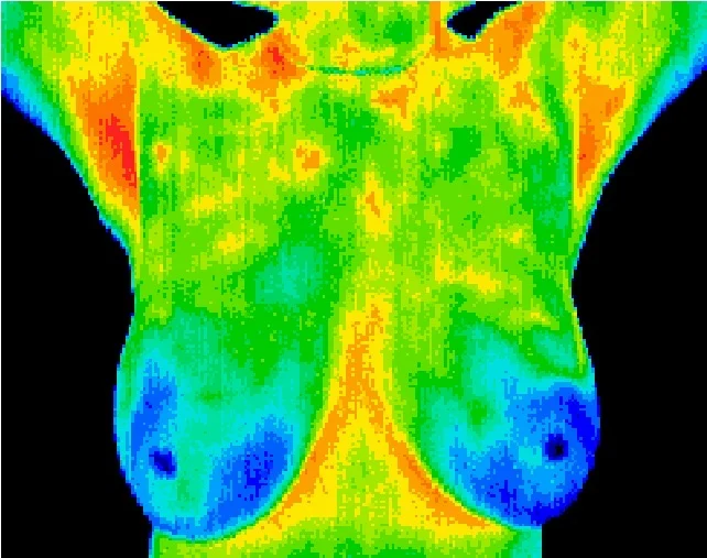
Breast thermal screening is a no-contact scan that records the pattern of heat across the chest wall. It is a functional picture, taken alongside the screening the NHS provides and the examination your GP does — never in place of either.
This is not a test for cancer.
Thermal imaging cannot detect, diagnose or rule out breast cancer, and it is not a replacement for a mammogram. That is the position of the MHRA here and the FDA in the United States, and it is the clinic’s position too. If you are eligible for NHS breast screening, go — and if you have already been invited and not been, that is the more useful appointment to make today.
If you have found a lump, or noticed any change — a change in shape or size, dimpling or puckering of the skin, a nipple that has turned inwards, discharge, or a rash or pain that is not going away — telephone your GP. Not us. Today, and not after a scan. Most changes turn out to be nothing serious, and the way to find that out is your GP.
What the scan actually does
A thermal camera photographs the heat your body gives off. Nothing enters the body, nothing touches it, and there is no X-ray dose and no compression — the camera simply records surface temperature and renders it as a colour map. It is the same equipment and the same physics as the thermal imaging used elsewhere in the clinic, pointed at a different part of the body.
What that map shows is physiology, not anatomy: blood flow at the surface, and how evenly it is distributed. Healthy tissue tends to be thermally symmetrical side to side, so the reading is largely a reading of asymmetry and of change over time — this side against that side, and this year against last.
It is worth being blunt about the limit of that. A temperature pattern is not a lump, a cyst or a tumour, and asymmetry has a long list of ordinary causes: hormones, where you are in your cycle, an old injury, a previous operation, even how you were sitting beforehand. A thermal study cannot separate those from anything else, and it is not being asked to.
Dr Jeffrey McTavish DC is a certified Level II Thermographer, which is the qualification governing how these images are captured and interpreted.
What it is used for
- Recording the pattern of surface heat across the chest wall and breast tissue
- Showing left-to-right thermal symmetry, and where it is not symmetrical
- Giving a comparable baseline, so the same person can be re-imaged and the two studies read side by side
- Adding a functional picture to an examination that is otherwise about structure
What it cannot do
- It cannot detect, diagnose or rule out breast cancer, or any other disease
- It cannot see inside the breast — it records surface temperature, and nothing else
- It is not a screening test, and it is not a replacement for mammography or ultrasound
- It cannot tell you a lump is harmless, and it is not the way to check one
Where it sits alongside NHS screening
Mammography and thermal imaging are not two versions of the same thing, and they are not in competition. A mammogram is a structural X-ray: it is looking for something physically present, and it is the test the national screening programme is built on because that is what the evidence supports. A thermal study is a functional picture of surface blood flow. One answers “is something there”; the other does not attempt to.
So the honest use of this scan is as something additional, for somebody who is already doing the things that matter: attending screening when invited, knowing how their own breasts normally look and feel, and taking any change to their GP. It is not a way to avoid a mammogram, and anyone who offers it to you as one is doing you harm.
Nothing found on a thermal study is a diagnosis. If a study raises anything the clinic thinks warrants a look, the answer is a letter to your GP, and the answer is the same whether or not you go on to have anything else done here.
What happens on the day
- You sit in a temperature-controlled room for around fifteen minutes first, so your skin settles. A scan taken straight in from the cold is not worth reading
- Nothing touches you and nothing is emitted. There is no compression, no radiation and no dye
- You undress to the waist for the imaging itself, and the camera is worked at a distance
- No deodorant, moisturiser or talc on the day; no hot shower, sauna, heavy exercise or physiotherapy in the four hours before
Ask the clinic whether a single baseline study or a baseline with a comparison scan afterwards suits you, and what it costs.
Tell us first
Nothing is emitted and nothing touches you, so there is no dose to weigh up and no reason the scan itself would be unsafe. What changes is whether it can be read: a great many ordinary things move surface temperature, and a study taken without knowing about them is a study nobody can interpret.
- Where you are in your cycle, and whether you are pregnant or breastfeeding
- Any breast surgery, implants, biopsy or radiotherapy, however long ago
- Hormone treatment, including HRT and the contraceptive pill
- Anything already under investigation — bring the letters, and tell us before the scan rather than after it
Questions before you book
Ask us whether it is worth it.
Sometimes the answer is that your GP or the screening programme is the right call and we are not — which is worth knowing before you pay for anything.
Breast thermal imaging is a functional imaging adjunct. It is not a screening test, not a diagnostic test, and not a substitute for mammography, ultrasound, clinical breast examination or the NHS Breast Screening Programme. Nothing on this page is a diagnosis and no outcome is promised. If you have a symptom, or you have been invited for screening, contact your GP or the screening service — they come first, and they come first whether or not you ever come here.
